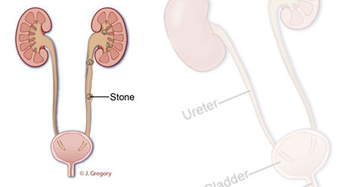
Nephrolithiasis is the presence of stones in the kidney; Urolithiasis is a more general term for stones anywhere within the urinary tract. This includes stones in the bladder, kidney, ureter (the tube leading from the kidney to the bladder), and urethra. Diagnosis is usually based on clinical history (sudden onset of excruciating pain in the flank along with blood in the urine, nausea or vomiting). The cause and chemical composition of the stone may have some bearing on its diagnosis, management and particularly on prevention of recurrence.
The peak incidence of stone disease occurs at the ages of 20-40 years, although stones are seen in all age groups. Males to female ratio is 3:1. Calcium oxalate stones, the most common variety, have a recurrence rate of 10% at 1 year, 35% at 3 years, and 50% at 5 years after the first episode. Kidney stones develop when crystals separate from the urine and aggregate within the urinary tract. Magnesium-ammonium-phosphate stones occur in the presence of infection. Uric acid stones form in patients with gout. Certain drugs and supplements, such as vitamin C, can also increase the risk of stones.
Most kidney stones pass within 48 hours with expectant treatment including hydration and analgesics. Ureteral stones less than 5 mm will pass spontaneously about 90% of the time.
For stones that do not pass or which are judged too large to pass spontaneously, a variety of procedures can be employed to remove the stone. No one technique is suitable for all stones in all patients, but the general options are as follows.
The least invasive procedure is called Extracorporeal Shock Wave Lithotripsy (ESWL). With this procedure, sound waves are focused on the stone. The sound then pulverizes the stone and the stone particles then pass through the urinary tract pushed along by the urine.
For stones that are too large, too hard or not easily seen with ESWL, two other procedures can be employed depending upon the location of the stone. For very large stones in the kidney, Percutaneous Nephrolithotripsy (PCNL) is employed. In this procedure, which requires general anesthesia and a short hospital stay, a tube is inserted directly into the kidney through the flank. The stone is then broken up and particles ‘vacuumed’ out of the kidney. As opposed to ESWL, there is no limit to the amount of power and energy that can be applied to the stone during PCNL, so even the hardest stones can be pulverized.
For smaller stones within the ureter and kidney, a procedure called ureteroscopy can be employed. In this procedure, performed under anesthesia on an outpatient basis, a special scope is used to navigate through the urethra, into the bladder and up the ureter. A laser fiber is then threaded through the telescope and brought into contact with the stone under direct vision. Fragmented particles are either removed or are allowed to pass spontaneously. Frequently, after the procedure, a thin silicone tube called a stent is inserted temporarily to allow any swelling in the ureter to subside. This is either removed by the patient several days after the procedure, or is removed by the doctor in the office.
More information:
Medicine.net | Kidney Stones
NKUDIC | National Kidney & Urological Diseases Information Clearinghouse


Urologists Dr. Steven M. Berman, MD and Dr. Mark Stein, MD specializing in Prostate, Kidney Stones, Erectile Dysfunction, Prostate Cancer, Kidney Cancer, Enlarged Prostate, Bladder Problems, Incontinence, and Female Urology.